The effectiveness of PIXCLARA in differentiating recurrent or progressive glioma from treatment-related change was evaluated in two clinical studies: a prospective, single center, open-label study (Study 1; NCT04044937), and a blinded central read study (Study 2) analyzing retrospectively collected images from multicenter, real-world clinical practice.
Study 1
Patients enrolled in Study 1 presented with intracranial neoplasm after primary treatment with radiation therapy, surgery, or both, and had suspected disease progression or recurrence based on MRI. The image analysis included a total of 127 patients (116 adults, 7 adolescents aged 12 years to 16 years, and 4 children aged 5 years to 11 years) with suspected recurrent or progressive glioma of any grade. Adult patients received 217 ± 23 MBq (5.9 ± 0.63 mCi) of PIXCLARA and pediatric patients received 155 ± 54 MBq (4.2 ± 1.4 mCi) followed by PET/MR imaging of the brain.
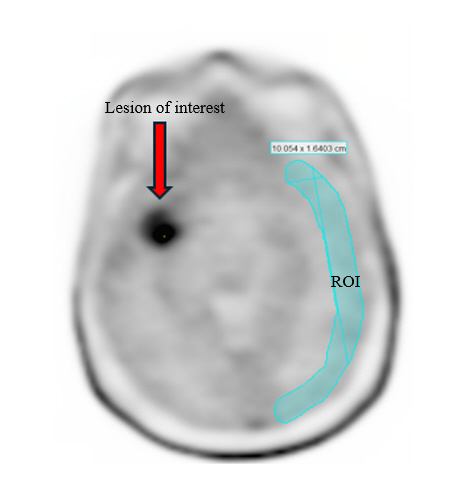
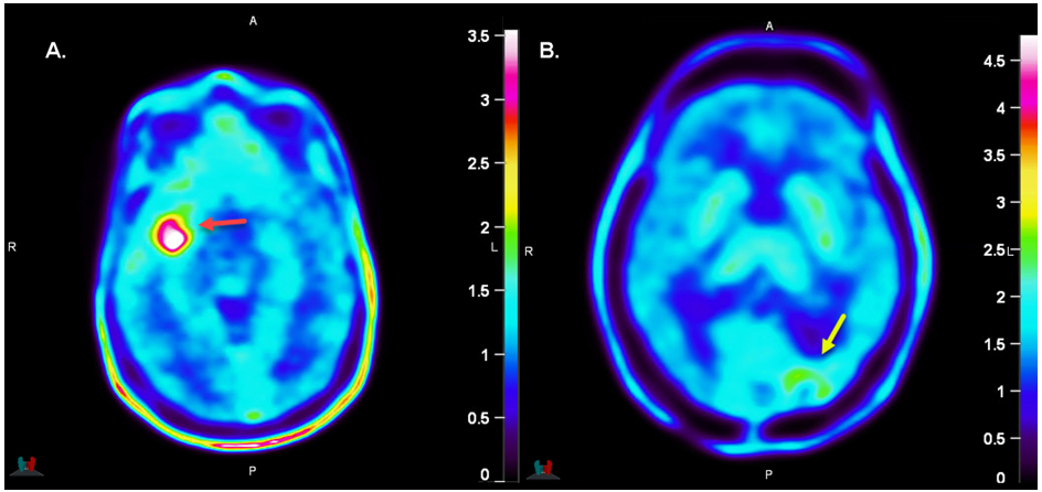
Three independent nuclear medicine experts performed blinded reads of the PIXCLARA PET images. Readers were blinded to all other clinical and imaging data except non-contrast T1 MR images used for localization of PET findings. Readers were instructed to identify up to five lesions and assign each as tumor (positive) or treatment-related change (negative) [see Dosage and Administration (2.5, 2.6)].
A multidisciplinary panel of experts determined the composite reference standard results. This panel reviewed any MR images obtained before, with, and up to 6 months after PIXCLARA PET, histopathology reports, additional conventional imaging reports, clinical reports, and surgical reports and assessed each lesion as tumor (positive) or treatment-related change (negative) based on totality of the available evidence. The panel did not have access to the blinded reader assessments or the PIXCLARA PET images.
The mean age of the patients in the efficacy analysis was 48 years (range 5 years to 85 years), and 62% were male. Of the study population, 73% were White, 14% Asian, <1% Black or African American, 12% of other or unknown race, and 8% Hispanic or Latino. Glioma grade was 1 (<1%), 2 (11%), 3 (21%), or 4 (67%). Isocitrate dehydrogenase was wild type in 60%, mutant in 31%, and not available in 9%. Bevacizumab was administered to 35% of patients within 6 months of PIXCLARA imaging. According to reference standard, 52% of patients had at least one lesion considered to be recurrent or progressive glioma.
PIXCLARA PET blinded reads were compared to the reference standard for detection of recurrent or progressive glioma at the lesion-level, and patient-level results, where at least one true positive lesion defined a true positive patient, are shown in Table 5.
Table 5: Lesion-Based, Patient-Level Performance of PIXCLARA PET for Detection of Recurrent or Progressive Glioma in Study 1 (n=127)For a given reader, the patient-level result is determined from the lesion-level result(s) using a composite reference standard.
| Endpoint | Reader 1 | Reader 2 | Reader 3 |
|---|
| Abbreviations: CI = confidence interval, n = number of patients, PPA = positive percent agreement, NPA = negative percent agreement |
| True Positive, n | 48 | 55 | 46 |
| True Negative, n | 36 | 29 | 39 |
| False Positive, n | 25 | 32 | 22 |
| False Negative, n | 18 | 11 | 20 |
| PPA PPA represents the percentage of patients with a positive PIXCLARA PET result among those who were reference standard positive. , %
(95% CI) | 73
(61, 82) | 83
(73, 90) | 70
(58, 79) |
| NPA NPA represents the percentage of patients with a negative PIXCLARA PET result among those who were reference standard negative. , %
(95% CI) | 59
(47, 71) | 48
(36, 60) | 64
(51, 75) |
Among the 11 pediatric patients included in the study, positive percent agreement (PPA) was 83% (95% confidence interval (CI): 44%, 97%) and negative percent agreement (NPA) ranged from 40% (95% CI: 12%, 77%) to 60% (95% CI: 23%, 88%) among the three readers.
Study 2
Patients included in Study 2 had known or suspected glioma of any grade treated with surgery, radiation, or chemotherapy, and had suspected disease recurrence or progression based on MRI. The image analysis included a total of 254 patients (253 adults and 1 adolescent aged 16 years). Adult patients received 201 ± 22 MBq (5.4 ± 0.58 mCi) of PIXCLARA and the pediatric patient received 181 MBq (4.9 mCi) followed by PET/CT or PET/MR imaging of the brain.
Three independent nuclear medicine experts performed blinded reads of the PIXCLARA PET images. Readers were blinded to all other clinical and imaging data except non-contrast T1 MR images used for localization of PET findings. Readers were instructed to identify up to five lesions and assign each as tumor (positive) or treatment-related change (negative) [see Dosage and Administration (2.5, 2.6)].
A multidisciplinary panel of experts determined the composite reference standard results. This panel reviewed any MR images obtained before, with, and after PIXCLARA, histopathology reports, clinical reports, and surgical reports and assessed each lesion as tumor (positive) or treatment-related change (negative) based on totality of the available evidence. The panel did not have access to the blinded reader assessments or the PIXCLARA PET images.
The mean age of the patients in the efficacy analysis was 51 years (range 16 years to 83 years), and 58% were male. Of the study population, 20% were White, 2% Asian, and 78% of other or unknown race. Glioma grade was 1 (<1%), 2 (17%), 3 (15%), or 4 (67%). Isocitrate dehydrogenase was wild type in 4%, mutant in 9%, and other or not available in 87%. Bevacizumab was administered to 4% of patients within 6 months of PIXCLARA imaging. According to the reference standard, 50% of patients had at least one lesion considered to be recurrent or progressive glioma.
PIXCLARA PET blinded reads were compared to the reference standard for detection of recurrent or progressive glioma at the lesion-level, and patient-level results, where at least one true positive lesion defined a true positive patient, are shown in Table 6.
Table 6: Lesion-Based, Patient-Level Performance of PIXCLARA PET for Detection of Recurrent or Progressive Glioma in Study 2 (n=254)For a given reader, the patient-level result is determined from the lesion-level result(s) using a composite reference standard. These results are consistent with those of a tipping point analysis using p=0, i.e., all missing PET are imputed as negative. The numbers of reference-positive and reference-negative are consistent across the readers.
| Endpoint | Reader 1 | Reader 2 | Reader 3 |
|---|
| Abbreviations: CI = confidence interval, n = number of patients, PPA = positive percent agreement, NPA = negative percent agreement |
| True Positive, n | 84 | 92 | 85 |
| True Negative, n | 77 | 67 | 72 |
| False Positive, n | 50 | 60 | 55 |
| False Negative, n | 43 | 35 | 42 |
| PPA PPA represents the percentage of patients with a positive PIXCLARA PET result among those who were reference standard positive. , %
(95% CI) | 66
(58, 74) | 72
(64, 79) | 67
(58, 75) |
| NPA NPA represents the percentage of patients with a negative PIXCLARA PET result among those who were reference standard negative. , %
(95% CI) | 61
(52, 69) | 53
(44, 61) | 57
(48, 65) |